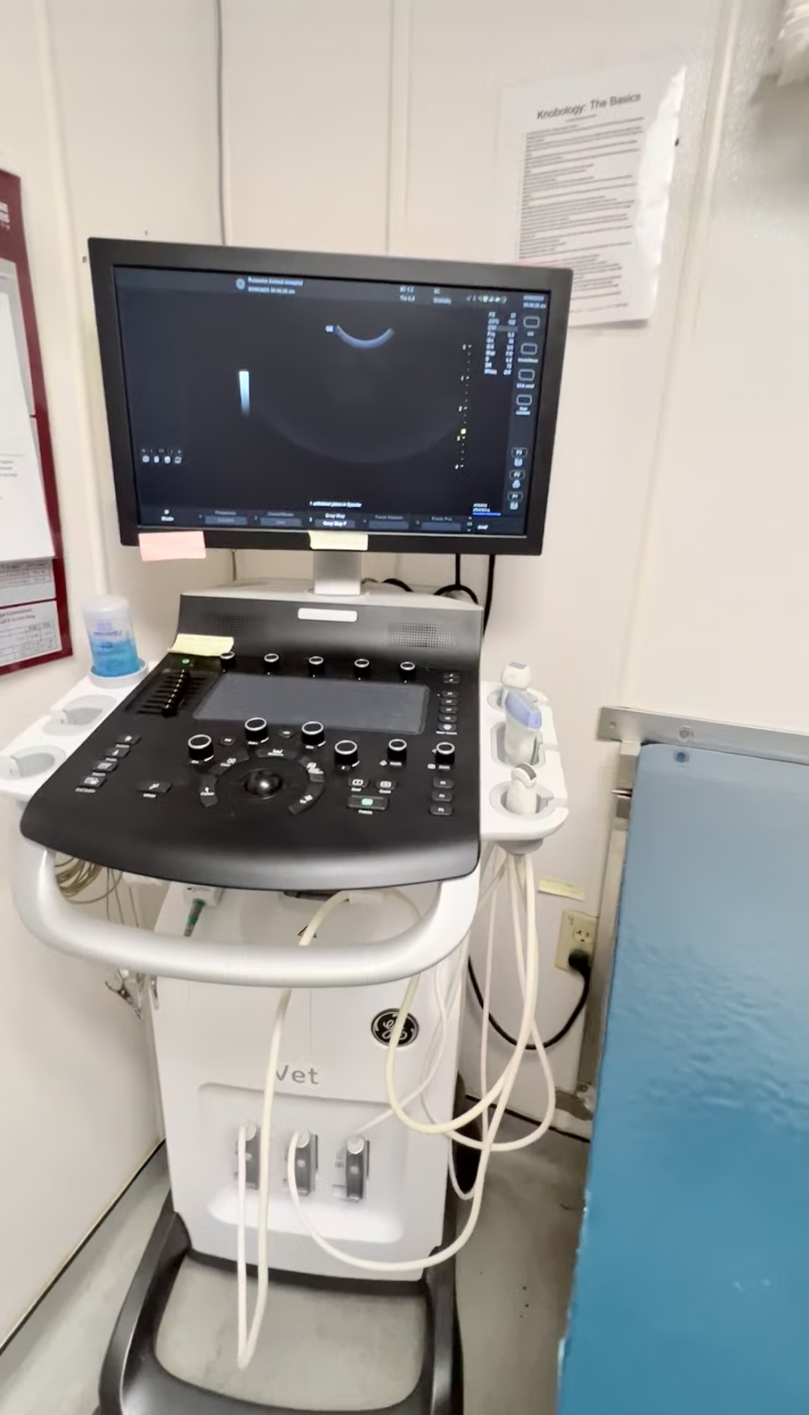
Ultrasound
our Ultrasound services
An ultrasound (also referred to as a sonogram) is a non-invasive, pain-free procedure used to evaluate the internal organs in pets without having to perform surgery. Ultrasound equipment directs a narrow beam of high-frequency sound waves into the area of interest. The sound waves may be transmitted through, reflected or absorbed by the tissues that they encounter.
The ultrasound waves that are reflected will return to the probe as “echoes,” and are converted into an image that is displayed on the monitor, giving a 2-dimensional real-time “picture” of the tissues under examination.
When Are Ultrasounds Used?
Ultrasound is invaluable for the examination of internal organs and was first used in veterinary medicine for pregnancy diagnosis. However, ultrasound has become extremely useful in evaluating the:
- abdominal organs
- certain structures in the neck
- the heart (echocardiogram)
- eyes
- muscles/tendons
- reproductive organs
For many abdominal disorders, both ultrasound and X-rays are recommended for optimal evaluation. The X-ray shows the size, shape and position of the abdominal contents, and the ultrasound allows the veterinarian to see inside the organs.
An abdominal ultrasound is indicated to evaluate pets with abdominal symptoms such as vomiting, diarrhea, straining to urinate or urinating blood. This test can also be helpful in cases of reproductive abnormalities, unexplained fever, loss of appetite or weight loss.
An abdominal ultrasound is often done if an X-ray, blood tests, or physical examination indicate a problem with an abdominal organ such as the liver, spleen, or pancreas. If physical examination reveals abdominal pain or enlargement of an abdominal organ, ultrasound examination could be indicated.
As with people, the abdominal ultrasound can also be used to detect early pregnancy and determine the viability of the fetus later in the pregnancy.
Ultrasound FAQs


